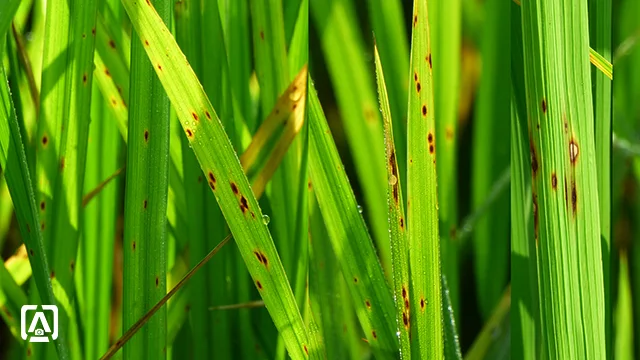
False smut of rice
Host: Paddy (Oryza sativa L.)
Pathogen: Ustilaginoidea virens (Cooke) Takahashi
Pathogen: Ustilaginoidea virens (Cooke) Takahashi
Distribution
False smut of rice is also known as green-smut or pseudo-smut. The
disease is prevalent in destructive form in major rice growing regions such as
China, India, USA. False smut of rice occurs as epidemics in India. It has been
reported from Andhra Pradesh, Bihar, Gujarat, Haryana, Jammu and Kashmir,
Jharkhand, Karnataka, Maharashtra, Pondicherry, Punjab, Tamil Nadu, Uttar
Pradesh and Uttaranchal of India (Muniraju et al., 2017). The disease causes
loss in yield as well as in quality. False smut of rice is so destructive that
it has caused up to 10.91 % loss in yield in Egypt (Atia, 2014). Intensive use
of fertilizer, cultivation of hybrid varieties and climate change are responsible
for the outbreak of the disease (Lu et a., 2009).
Symptoms
 |
| Grain of rice transformed in to smut ball |
Symptoms appear in
the form of smut balls. These are transformed rice grains. Only a few grains in a
panicle are affected. However, in severe cases, all the grains in a panicle are affected (personal observation since 2013-2020). Developing smut balls are small flattened and covered by a
thin membrane. At maturity, growth of fungus is intensified and smut ball bursts
to disperse the chlamydospores. As chlamydospores develop, smut balls become
orange colored, which later on become yellowish-green color and finally turned greenish-black
colored.
 |
| Heavily infected panicles of rice showing smut grains |
Pathogen
 |
| Spores of Ustilaginoidea virens |
False smut of rice is caused by Ustilaginoidea virens. The
perfect stage of this fungus is Villosiclava virens. In potato-sucrose agar medium it produced slow-growing colonies. Conidia are elliptical (measuring
3-5 µm in diameter) which on maturation or under unfavorable condition, develop into round spinulate
chlamydospores (Wang et al., 2019). Wang et al. (2019) reported one or two horse-shoe shaped sclerotia development in smut balls.
Annual recurrence and Disease cycle
 |
| A "combine machine" harvesting the rice from the "false smut of rice" infected farm. A cloud of chlamydospores are blown in the air which, through the wind dispersed at a large distance. |
The disease is soil and seed born. In the plots where mechanized harvesting is practiced through combine the machine, a cloud of chlamydospores are blown into the atmosphere and spread in a large areas. Finally, these spores are settled-down in the soils and stubble, where they survive in the soils for a longer time. On getting susceptible host in the coming season, chlamydospores germinate infect the paddy through the roots.
Olive green to brown colored fruiting bodies so-called "sclerotia" are produced in the smut balls. Sclerotia settle down on the soils and overwinter. On getting favorable conditions, sclerotia germinate into the stalked structures bearing perithecia. These perithecia are flask-shaped structures bearing ascus. Ascus produced filiform ascospores, which liberate and germinate to start a new cycle.
 |
| Disease cycle and annual recurrence of false smut of rice |
Olive green to brown colored fruiting bodies so-called "sclerotia" are produced in the smut balls. Sclerotia settle down on the soils and overwinter. On getting favorable conditions, sclerotia germinate into the stalked structures bearing perithecia. These perithecia are flask-shaped structures bearing ascus. Ascus produced filiform ascospores, which liberate and germinate to start a new cycle.
Control measures
- Early-planted rice are less affected
- Seed should be treated with carbendazim @ 2.0 g/kg seed.
- Excess nitrogenous fertilizers should be avoided.
- Spray of Hexaconazole @ 1ml/liter reduced the infection.
- Spray of Mancozeb or carbendazim or copper-oxychloride inhibit the conidial germination and secondary infection.
- Stubble should be removed from field and burnt.
Sources
- Atia, M.M., 2004. Rice false smut (Ustilaginoidea virens) in Egypt. Journal of Plant Diseases and Protection, 111(1), pp.71-82.
- Lu, D.H., Yang, X.Q., Mao, J.H., Ye, H.L., Wang, P., Chen, Y.P., He, Z.Q. and Chen, F., 2009. Characterising the Pathogenicity Diversity of Ustilaginoidea Virens in Hybrid Rice in China. Journal of Plant Pathology, pp.443-451.
- Muniraju, K.M., Pramesh, D., Mallesh, S.B., Mallikarjun, K. and Guruprasad, G.S., 2017. Novel fungicides for the management of false smut disesases of rice caused by Ustilaginoidea virens. Int J Curr Microbiol App Sci, 6(11), pp.2664-2669.
- Wang, W.M., Fan, J. and Jeyakumar, J.M.J., 2019. Rice False Smut: An Increasing Threat to Grain Yield and Quality. In Protecting Rice Grains in the Post-Genomic Era. IntechOpen.
Brown spot of rice
Host: Paddy (Oryza sativa L.)
Pathogen: Helminthosporium oryzae (telomorph: Cochliobolus rniyabeanus)
Pathogen: Helminthosporium oryzae (telomorph: Cochliobolus rniyabeanus)
Distribution
Brown spot of rice is reported from many rice growing countries of the world, including India, Nepal, Philippines, Surinam, Sumatra and Sierra Leone. This disease can cause up to 90 % yield loss in rice.
This disease is historically important as it has caused two devastating epidemics in India; one during 1918-19 in the Krishna-Godavari delta in the southern part and another in 1942-43 in Bengal. The latter is famous as the Bengal famine of 1943.
This disease is historically important as it has caused two devastating epidemics in India; one during 1918-19 in the Krishna-Godavari delta in the southern part and another in 1942-43 in Bengal. The latter is famous as the Bengal famine of 1943.
Symptoms
 |
| Infected rice leaves showing brown spot |
Typical brown to grayish colored spots appear on the aerial part of the rice plant, including coleoptiles, leaf sheaths, leaf blades and glumes. The spots are ellipsoidal to oval in shape with grayish-brown in the center and bright reddish-brown margin. In severe cases, grains lose bright color and grain is shriveled, so called "pecky rice".
 |
| Typical brown spots on rice leaf |
Pathogen
The causal organism of brown spot of rice is recognized as the fungus, Helminthosporiurn oryzae Breda de Haan. The perfect stage (telomorph) of this fungus is described as Cochliobolus miyabeanus.
Control measures
Disease is soil as well as seed-born. The pathogen survives in the infected grains for up to the duration of 4 years.
- Destroy the debris leftover after harvesting to kill the fungus from the field.
- Certified and disease-free seed should be used.
- In order to prevent the disease incidence, seed should be treated in fungicides such as, carbendazim, propiconazole, azoxystrobin.
- Very dense plantations should be avoided to reduce the humidity.
- Excess nitrogenous fertilizers should be avoided.
- Resistant varieties of rice should be sown.
Sources
- Chakrabarti, N.K., 2001. Epidemiology and disease management of brown spot of rice in India. In Major Fungal Diseases of Rice (pp. 293-306). Springer, Dordrecht. (https://link.springer.com/chapter/10.1007/978-94-017-2157-8_21).
Stem borer of rice
Host: Paddy (Oryza sativa L.)
Pathogen:Yellow stem borer insect (Scirpophaga incertulas Walker)
Pathogen:Yellow stem borer insect (Scirpophaga incertulas Walker)
Distribution
Yellow stem borer is one of the destructive pests of rice distributed throughout the world including India, Pakistan, Nepal, Sri Lanka, Bangladesh, Afghanistan, Indonesia, Burma, Malaysia, China, Taiwan, Japan, Philippines, Singapore, Thailand, Tanzania, Vietnam, Iraq, Brunei, etc. In India yellow stem borer is distributed in rice growing states and causes yield loss of 20 to 80 %.
Symptoms
"Whiteheads" of panicles, which are unfilled and empty are typical symptoms of the yellow stem borer infestation. Larvae of the insect feed upon the leaves leaving white-yellow longitudinal patches on the leaves and leaf sheath as well. The larvae bore the hole in the parenchyma of the hollow stem near the node and enter in the pith, where they feed upon the stems from inside. This causes damage to the stem and infested panicles of rice can be easily pulled-out from the plant. Damage caused by the larvae of insect cuts the nutrient supply to the heads. As a result the whole panicle becomes white, unfilled and remains empty.
The infested and bored stems give characteristic foul odor.
Upon splitting of infested stems, brown color egg mass and faecal matter of larvae can be easily seen.
The infested and bored stems give characteristic foul odor.
Upon splitting of infested stems, brown color egg mass and faecal matter of larvae can be easily seen.
Pathogen
 |
| Larvae of stem borer of rice inside the hollow pith of rice stem (above) and round holes bored by the larva on the stem (below). Scale = 1.0 mm. |
Yellow stem borer is a moth belonging to the family Crambidae of Lepidoptera. The moth is of small size ranging between 12 to 34 mm wing span. Male adults are smaller than female one. Numerous eggs in masses are laid-down by the adult female. Within a week eggs hatch out into pale yellow larvae ranging between 4 to 6 mm in length. Larvae after feeding 2 to 3 day in the leaf sheath, enter the stem near the node, where they develop into the pupa. The pupa finally develop into the adult moth.
Control measures
Always adapt the Integrated Disease or Pest Management (IPM) to control this disease.
- Late plantation favors the yellow stem borer. So, early rice plantations should be practiced.
- Avoid close plantation.
- Remove the infection plants manually at the deadheart stage only.
- Use pheromone traps and yellow sticky traps to catch the moths.
- Use resistant varieties only.
- Prune the tip of leaves containing eggs after transplantation of nursery.
- Avoid excess use of nitrogenous fertilizers.
- Remove and destroy stubbles at the end of the crop.
- Release natural predators and parasitoids, e.g., Trichogramma japonicum starting after 15 days of transplantation, which eat upon the eggs and larvae of moth.
- Infestation can be prevented by dipping the roots of seedlings for 12 to 14 hours in the 0.02 % chlorpyrifos.
- 0.02 % chlorpyrifos should be sprayed once the population of male adults reaches 25 to 30 per trap per week.
Potassium deficiency in rice
Symptoms
 |
| Rice leaves showing browning at the tip and margin |
Rice plants deficient in K show characteristics symptoms shown in above figure. First of all, the tip of lower leaves turn yellow which gradually grow along both the margins towards the base of leaves. Soon the yellow portion turns scorchy. If not treated, entire leaves become brown and scorchy and eventually fall.
Management
K-deficiency is treated by the application of potassium-containing fertilizers. There are various fertilizers containing potassium. These are
Among these , potassium chloride (KCl), also known as muriate of potash (MOP) is the most commonly available and widely used potassium fertilizer. It contains approximately 60 to 62 % K2O (50 % K). Iron impurities present in the muriate of potash gives the red-color appearance.
- Potassium chloride
- Potassium nitrate
- Monopotassium phosphate
- Potassium sulfate
- Potassium thiosulfate
Among these , potassium chloride (KCl), also known as muriate of potash (MOP) is the most commonly available and widely used potassium fertilizer. It contains approximately 60 to 62 % K2O (50 % K). Iron impurities present in the muriate of potash gives the red-color appearance.
Dose of fertilizer
Depending upon the target yield, application of 10 to 20 Kg muriate of potash per acre cures the K-deficiency.
Content first created on 30-08-2020
last updated on 26-09-2024
last updated on 26-09-2024


0 Comments
Leave your comments here.